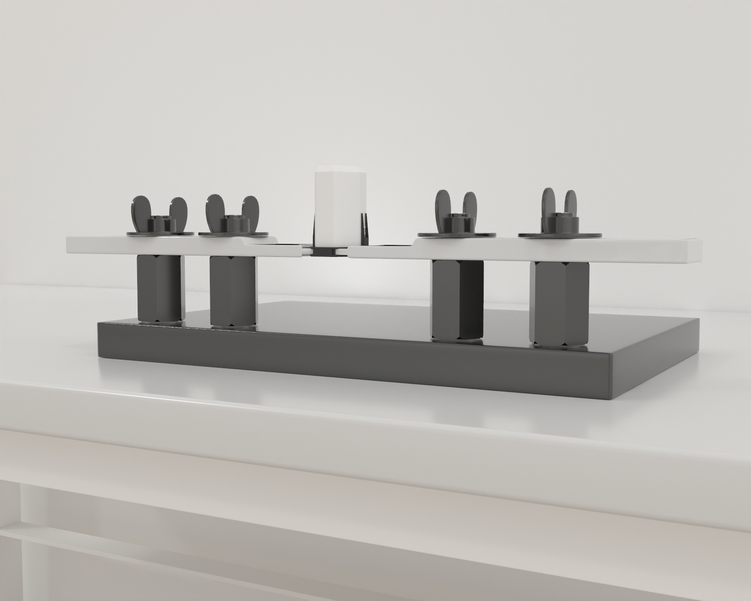
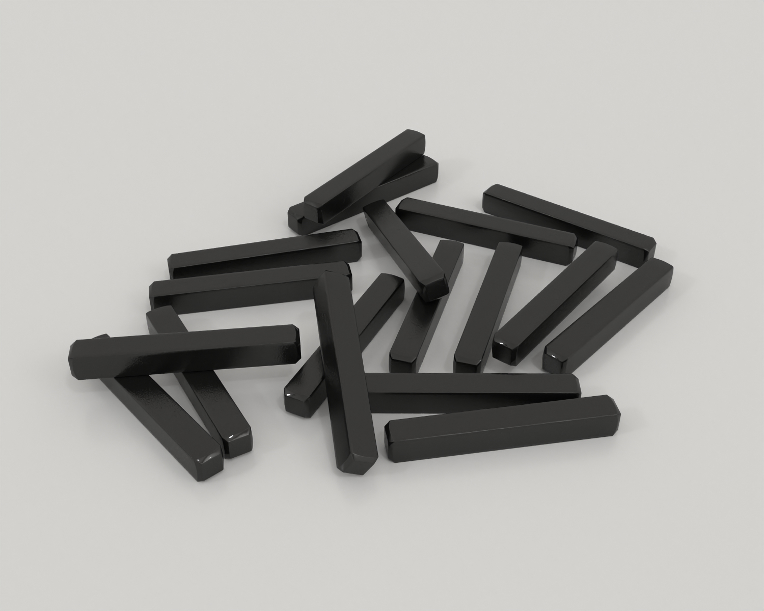
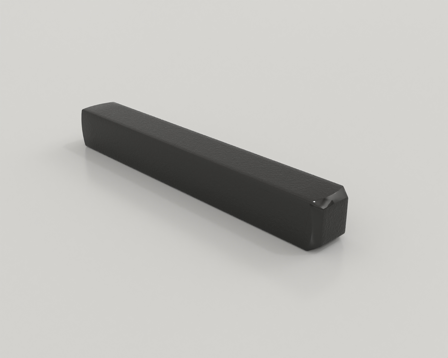
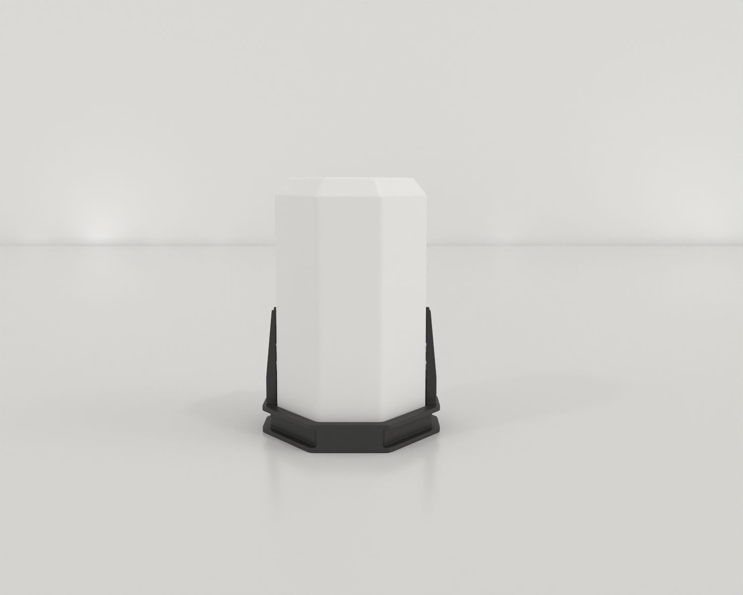
The head bar and head hat apparatus are to be used with the Maze Engineers Head Fixation Station apparatus. The head bar is designed to be as less intrusive than other traditional and commercially available designs while the modular head hat design protects head- and skull-mounted hardware. These apparatuses can be implanted using stereotaxic surgical equipment.

Both the head bar and head hat dimensions and specifications are customizable, adjustable, and scalable to each laboratorys needs.

Head fixation devices are commonly used for studying anesthesia effects, facial function, neuroimaging, reflex adaptation, operant conditioning, and reflexes such as eye blinking in mice (Schwarz et al., 2010).

Price and Specifications

Mouse Head Bar

$ 200

Per Month
  • Sizes available: 20mm-40mm  Length X  3.2 Width X 3.2 mm Height
  • Weight 1.1g
  • Composed of Titanium
  • Please include the size required when requesting a quote (customizable sizing available)
  • Customizable sizing available in 1mm increments
  • 25 units

Rat Head Bar

$ 250

Per Month
  • Sizes available: 40-200mm L in 1mm increments x
  • Composed of Titanium
  • Customizable sizing available in 1mm increments
  • Please include the size required when requesting a quote
  • 25 units

Introduction

Head fixation is a common procedure used in neurological studies. The main purpose of head fixation devices is to restrict the subject movement. Apart from motor reflexes, as described earlier, the head fixation device can be utilized in a neuroscience lab for training mice in operant conditioning tasks. In addition, head-fixed mice can be trained to differentiate between auditory, visual, and olfactory stimuli.

The head bar affixes the subject to the head fixation station while the headhat protects head-affixed hardware (such as implanted electrodes and cannulas) from damage. Both the head hat and head bars are customizable in sizing, see our pricing table for default sizing.

The head bar is necessary for use of the head fixation station to impact head and eye movement with respect to visual stimuli. These shapes can be altered in dimension to fit your subjects.

The head hat system is optional for the use and protection of skull- or head-affixed apparatus such as cannulas, electrodes, optogenetic hardware etc. We offer two sizes of head hats, a compact design for cannula guides and smaller skull-affixed apparatus or a larger design to provide greater protection for a greater number of probes.

Head Fixation Protocols

Before beginning this procedure, ensure your methods and lab adhere to laboratory animal safety regulations applicable to your country and institution.

  1. During surgery on a stereotaxic apparatus, while the subject is under anesthesia, cut away all tissue from the skull below the desired attachment location of the headbar. Score the skull with a scalpel for good adhesion. Perform the intended surgery (implant, injection, etc). Ensure that the headbar will fit over the top of any implant. If this is not the case, be sure to place the headbar before the implant.
  2. Coat the exposed area of the skull with Optibond and allow it to dry. This creates a prepared surface to which later adhesives can form a secure attachment to. Sterilize the headbar by quickly submerging in ethanol and allow to dry.
  3. Coat the bottom of the headbar in cyano-acrylate adhesive and place the headbar on the skull in the desired location with the clip attach points along the medial–lateral axis. The adhesive should hold within a few minutes.
  4. Assuming all implants are in place and the surgery is at its end, fill the cavity between the skull and the headbar to the ledge with dental cement and let it cure. Make sure not to overfill this cavity as the headhat attachment points must remain clear. Following the surgery, let the subject recover.
  5. When the subject has recovered and the glue is dry, align the headhat to the headbar, and press down to secure. The clip mechanism should engage and feel secure against the headbar. To release the headhat, press in on the side tabs and the headhat should come right off.

Rodent behavior and movement can be tracked using a video tracking software package such as Noldus EthoVision, ANY-Maze, or BehaviorCloud.


Data Analysis

The following parameters can be observed using the Octopus three-chambered social apparatus:

Literature Review

An open-source head-fixation and implant-protection system for mice

The authors introduce an available solution for securely immobilizing the heads of mice during neuroscientific experiments. The system is designed to minimize discomfort for the animals while ensuring the stability of implanted devices.

The paper presents a detailed description of the hardware components of the system. The system incorporates a custom 3D-printed head-fixation module, which allows for precise adjustment and stabilization of the mouse’s head position.

One key feature of the system is the implementation of an implant-protection mechanism. It employs a transparent protective cap that covers the implanted devices, shielding them from potential damage caused by the rodent’s movements. This safeguarding measure increases the longevity and reliability of the implants, enabling long-term experiments.

Overall, “An open-source head-fixation and implant-protection system for mice” provides practical solutions that address the challenges associated with head-fixation and implant protection in mouse experiments.

Summary

  • The rodent head bar and hat are used in conjunction with the rodent head fixation system.
  • The rodent head bar affixes the rodent skull to the fixation system.
  • The rodent head hat protects cannulas, wires, and apparatus while using the fixation system.

References

Weaver, I. A., Aryana Yousefzadeh, S., & Tadross, M. R. (2023). An open-source head-fixation and implant-protection system for mice. HardwareX13e00391. https://doi.org/10.1016/j.ohx.2022.e00391

Are you an academic scientist and creator?

Learn how your lab can tech transfer this and similar devices from your lab to industry.