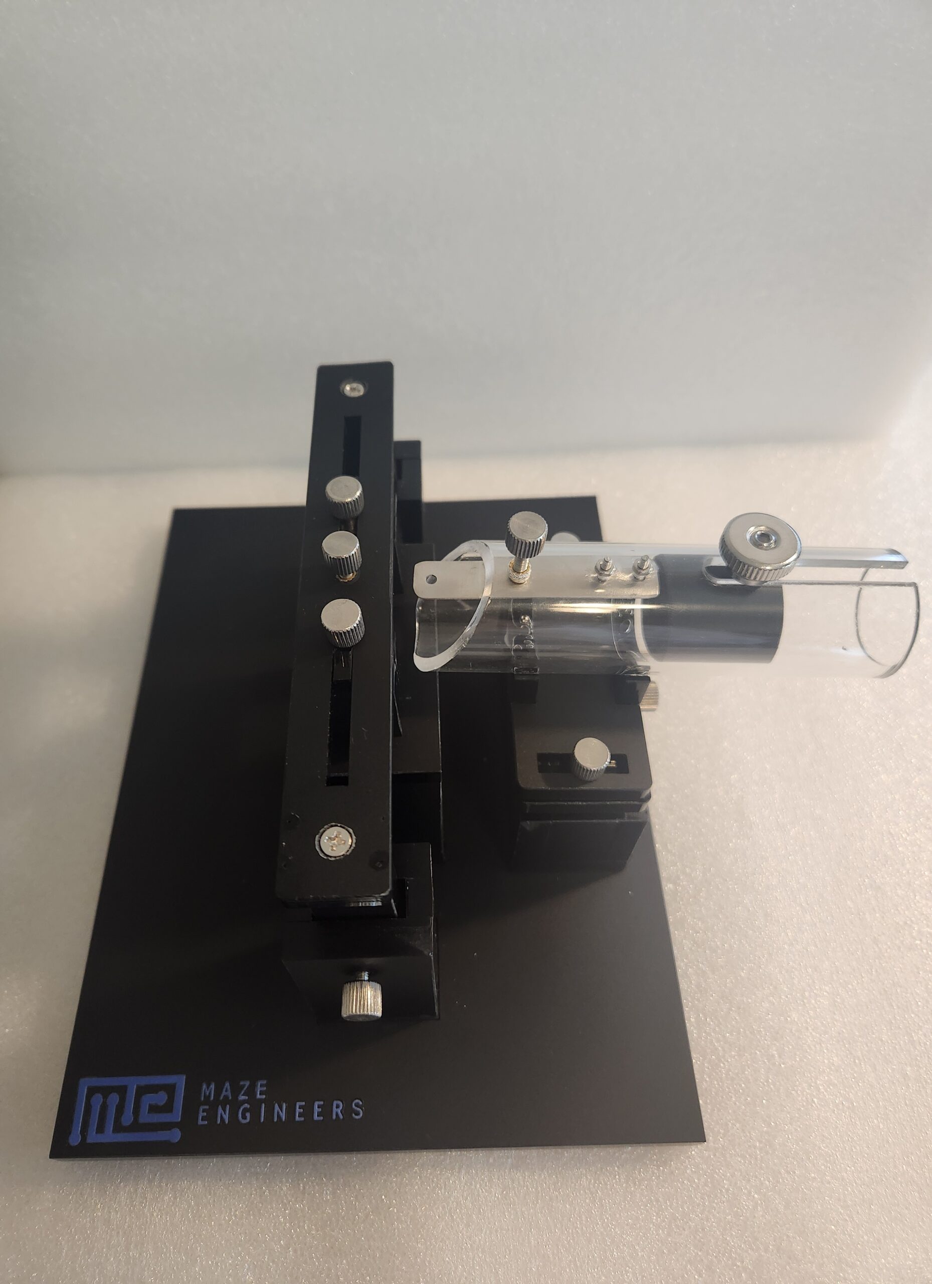
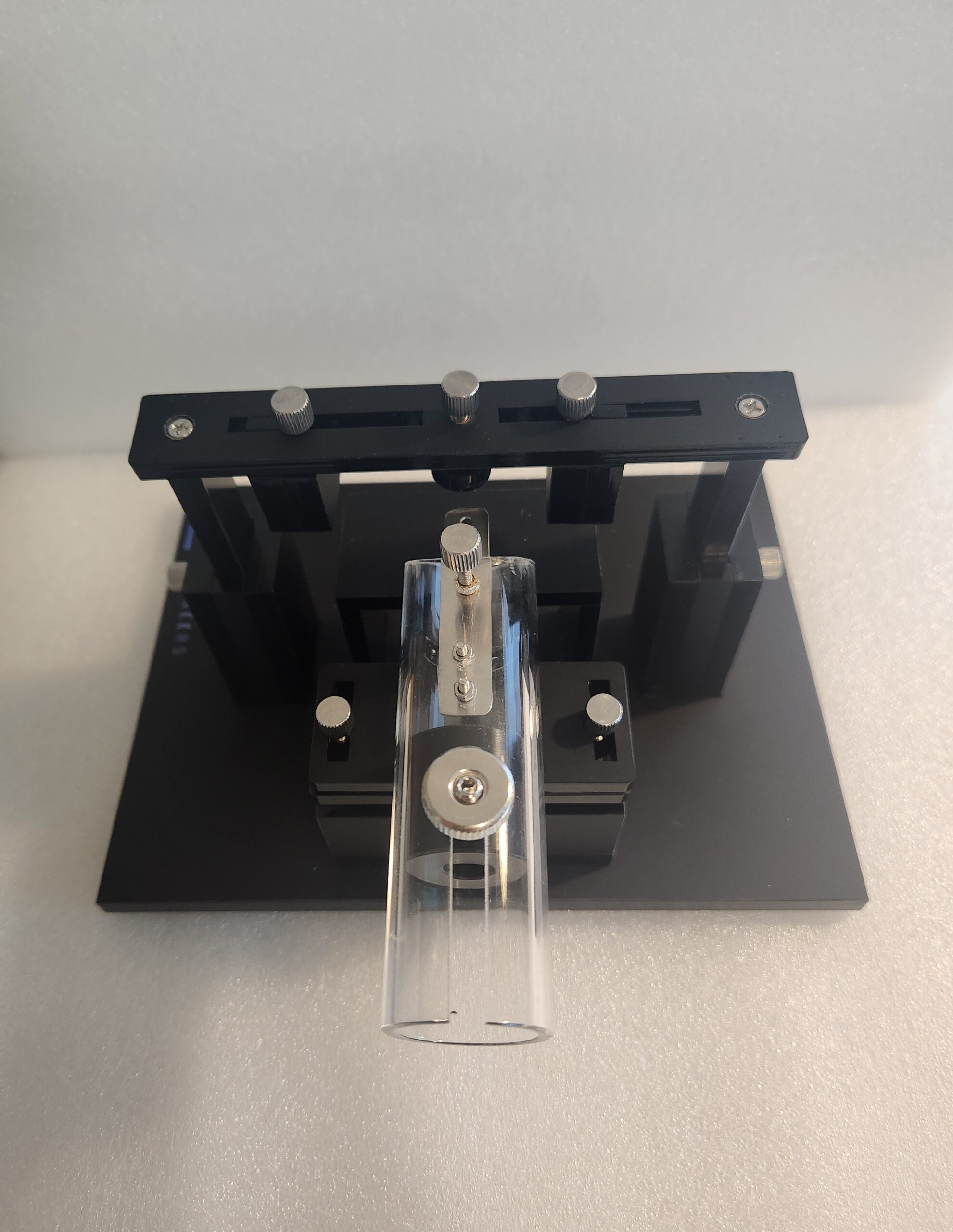
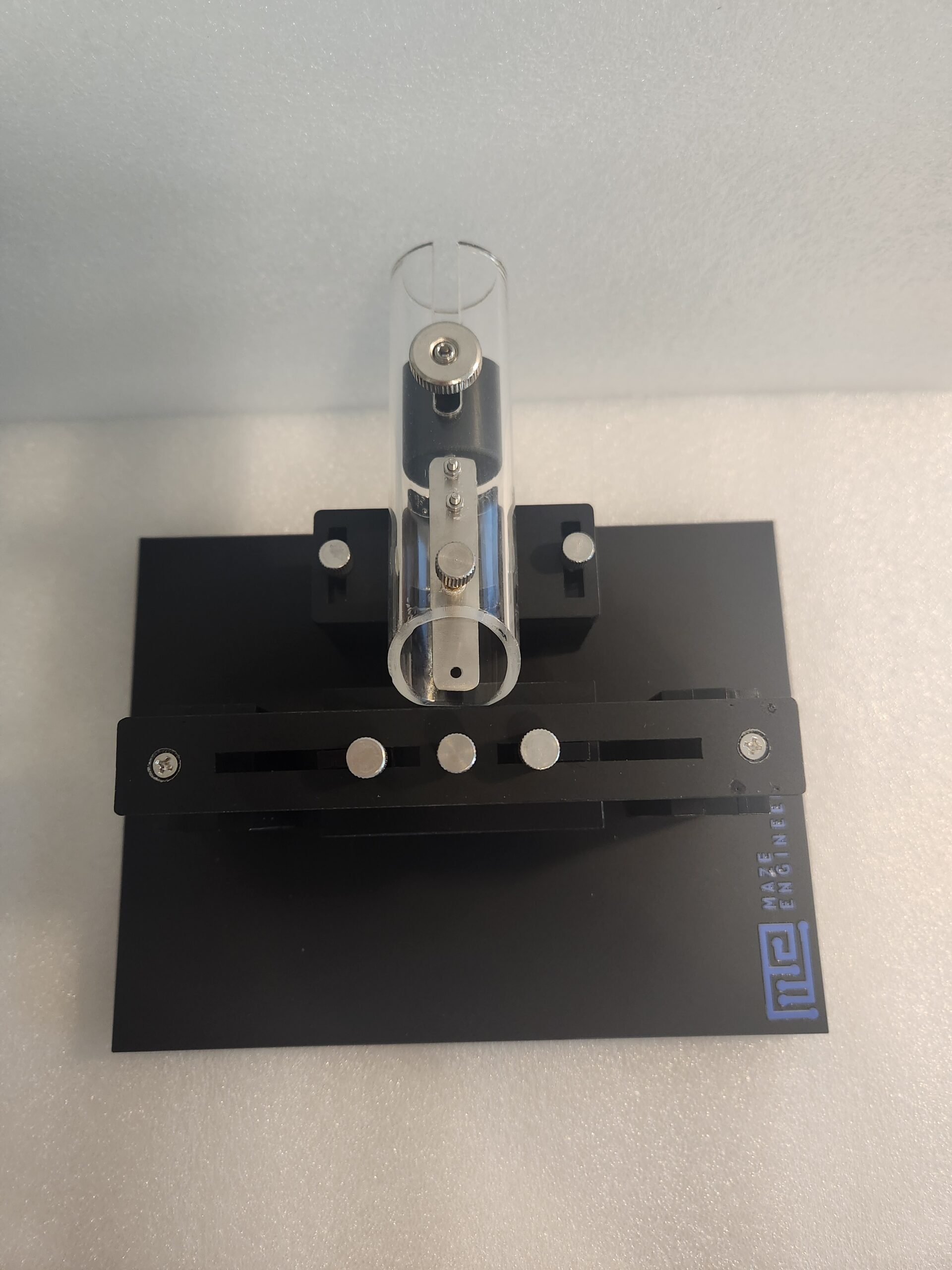
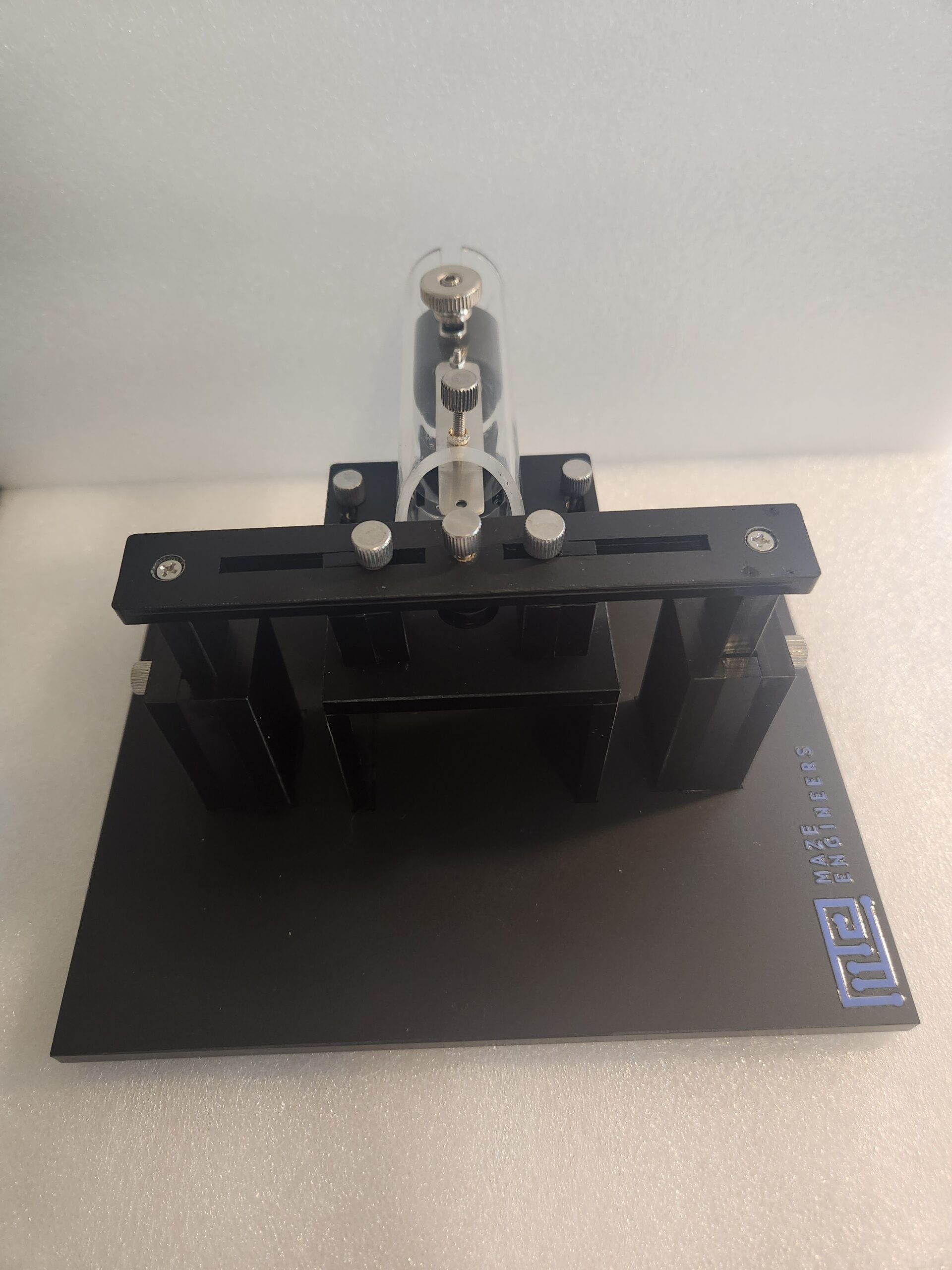
The Maze Engineers Head fixation is employed in neurobiological laboratories to restrain murine head movement during a multitude of behavioral paradigms. A head fixation system comprises thumbscrews that can be connected to the head bars, and a clear acrylic tube to hold the animal during the experiment.

Head fixation devices are commonly used for studying anesthesia effects, facial function, neuroimaging, reflex adaptation, operant conditioning, and reflexes such as eye blinking in mice (Schwarz et al., 2010).

Price and Specifications

Mouse

$ 325

Per MonthHead fixation system:

  • Acrylic introduction tube: 1.5 inches
  • Aluminium headboard: 150mm x 150mm x 12.7 mm

Rat

$ 425

Per MonthHead fixation system:

  • Acrylic introduction tube: 2.4 inches
  • Aluminium headboard: 240mm x 240mm x 20.32 mm

Introduction

Head fixation is a common procedure used in neurological studies. The main purpose of head fixation devices is to restrict the subject movement. In addition, it enables the researchers to optically assess neuronal circuits thereby reducing background noise and motion artifacts. The head fixation system restrains the degrees of behavioral freedom. These include learning behaviors associated with reward and those involving defined motor skills such as eye movements, lever pulling, licking, and virtual reality (Weaver et al., 2023). In addition, head fixation devices are frequently employed for recording neuronal activity by using calcium imaging and electrophysiology techniques.

The head fixation process is crucial as it impacts to head and eye movement with respect to visual stimuli. Apart from motor reflexes, as described earlier, a head fixation device can be utilized in a neuroscience lab for training mice in operant conditioning tasks. In addition, head-fixed mice can be trained to differentiate between auditory, visual, and olfactory stimuli.

The head fixation process requires the attachment of the head bar to the animal’s head, immobilization of the head bar along with the subject’s head using an external apparatus, and a head hat to protect skull-mounted hardware.

Apparatus and Equipment

The head fixation system comprises a clear acrylic tube into which the subject is introduced. This head restrainer is designed to offer exceptional stability and comfort to the subject. The thumbscrews can be simply used to capture or release the animal with the help of titanium head bars (purchased here).

The head restrainer is pre-mounted on a metric aluminum headboard. Other specifications include 10mm vertical travel to maximize animal comfort, autoclavable material, and the ability to be used with other existing experimental setups. Mazeengineers offer a head fixation system with an ergonomic design.

Data Analysis

The following can be studied using the Rodent Head-Fixation apparatus:

  • Drug and anesthesia effects
  • Rodent psychophysics
  • Rodent fine motor skills such as facial function eg. blinking
  • Motor skills such as reflex adaptions and actions
  • Neural and biomechanical force production
  • Neuroimaging techniques such as Optogenetics
  • Electrophysiology
  • Operant conditioning

Protocol

(Guo et al., 2014)

Head Bar Implantation

  1.     Deeply anesthetize the mice using 2% isoflurane and mount the subject on the stereotaxic frame.
  2.     Place the mice on a heating pad and cover its eyes in a thin layer of petroleum jelly.
  3.     Clean the subject’s scalp with 70% ethanol and betadine and inject 50ꭒl 0.5% Marcaine under the scalp for topical anesthesia. In addition, inject an anti-inflammatory drug like ketofen subcutaneously and buprenorphine intraperitoneally.
  4.     Following this, remove approximately 1cm2 flap of skin from the dorsal skull with a single cut and clean the remaining gelatinous periosteum with small scissors. Then clean and dry the skull using a sterile cotton swab.
  5.     Expose the skull by scrapping off the bone with a scalpel and cover the exposed skull with a layer of cyanoacrylate glue.
  6.     Place the head bar directly into the wet glue. Use dental cement to cover the glue and secure the head bars in place.

Head-fixation Surgery

  1.     Place the wings of the head bar into the notches in a stainless steel holder using a pair of thumbscrews and clamps.
  2.     Now, insert the mouse body into the acrylic body tube such that the mouse’s head extends out and its front paws grip the tube’s edge after head fixation.
  3.     After that, attach the holder along with the body tube to a caddy. The head bar should be 30mm above the body tube’s bottom. Fix the caddy into the behavior box. Always remember that a head-fixed mouse should crouch in the body tube in a natural position.
  4.     Following head fixation, the mice can be subjected to various experiments by using accessories like lickport to study the neurobiology of rodent behavior.

Literature Review

Monitoring of Force generated during Behavior

Hughes et al. (2020) tried studying the subtle movement and postural adjustments that animals experience during head fixation. The experimenters utilized a novel mouse head fixation system equipped with five orthogonal force sensors to study in vivo electrophysiology of the mouse brain during behavioral tasks like licking.

They housed the mice in groups of 3-4 mice per cage following a 12:12 light cycle and conducted the experiments in the light phase. The subjects were deprived of water and allowed free access to water for about 2h after the experimental sessions. The scientists attached the mouse perch and head fixation apparatus to a base plate made of steel and elevated this assembly to accommodate the reward delivery apparatus. The load cells were also added to the experimental setup. Following this, the researchers anesthetized the mice with 2-3% isoflurane and performed craniotomy using a stereotaxic frame. During the surgery, they implanted 16 electrodes in a 4×4 configuration using thumbscrews and secured them using dental acrylic. Then, they also inserted a metal bar and cemented it using dental acrylic.

Ultimately, the experimenters trained the mice for a Pavlovian conditioning task and observed the licking behavior. They concluded that licking-related oscillations revealed postural alterations among the subjects.

Strengths and Limitations

Precautions

  •  The animals chosen for head-cap implantation using a head fixation system should be optimally 12 weeks of age. By this time, the females are fully grown up whereas males can grow for a longer time.
  • Make sure that the animal becomes acquainted with the researcher before conducting the behavioral task. For this, simply place your hand in the cage and let the animal explore it.
  • If you’re using a cream for depilation, make sure to properly rinse it afterwards.
  •  During stereotaxic surgery using a head fixation device, use a heating pad to maintain the animal’s body temperature at 37oC (Schwarz et al., 2010).
  •  Try employing single-housing mice to prevent the damage caused to implants by cages.

Strengths and Limitations

The head fixation system is an efficient device with an ergonomic design used in neurobiology labs to affix murine heads. The foremost advantage of head-fixation devices is they facilitate easy monitoring of murine neuronal activity thereby minimizing background noise and motion artifacts. The mechanical stability ensured by head fixation provides favorable conditions for visualizing neural activity in awake animals.  Secondly, the head fixation devices confer the advantage of greater experimental control to the researcher. Thirdly, even if the mice are not trained for a particular task, the electrophysiological neural signals are not affected by anesthetization in head-fixed awake mice.  In addition, modern head fixation devices offer a reduced footprint in the subject head (Schwarz et al., 2010).

However, there are a few disadvantages as well. For instance, the head-fixed behaving rat requires a lot of time to get habituated to the experimental setup. In addition, the animal is restricted to performing several whole-body movements under head fixation. Lastly, the head bars that are wider than the subject’s body can take longer than desired time to head fix the animal.  

Summary

  A head fixation system is employed in neurobiology laboratories to affix mouse heads during several behavioral paradigms.

  •       A head fixation system comprises a head restrainer pre-mounted on a metallic breadboard.
  •       It also contains thumbscrews which can be connected to titanium head bars (purchased separately), and a clear acrylic tube to hold the animal during the surgery.
  •       Head fixation devices are commonly used for studying anesthesia effects, facial function, neuroimaging, reflex adaptation, operant conditioning, and reflexes such as eye blinking in mice.

References

Guo, Z. V., Hires, S. A., Li, N., O’Connor, D. H., Komiyama, T., Ophir, E., … & Svoboda, K. (2014). Procedures for Behavioral Experiments in Head-Fixed Mice. PLoS ONE, 9(2).

Hughes, R. N., Bakhurin, K. I., Barter, J. W., Zhang, J., & Yin, H. H. (2020). A head-fixation system for continuous monitoring of force generated during behavior. Frontiers in integrative neuroscience, 14, 11.

Schwarz, C., Hentschke, H., Butovas, S., Haiss, F., Stüttgen, M. C., Gerdjikov, T. V., … & Waiblinger, C. (2010). The head-fixed behaving rat—procedures and pitfalls. Somatosensory & motor research, 27(4), 131-148.

Weaver, I. A., Yousefzadeh, S. A., & Tadross, M. R. (2023). An open-source head-fixation and implant-protection system for mice. HardwareX, 13, e00391.