


The Visual X maze or Visual-Stimuli 4-arm Maze (ViS4M), is used to quantitatively study visual and cognitive abilities in rodents. This apparatus can be used to assess disorders such as early functional decline associated with aging and Alzheimer’s disease progression (Vit et al., 2021, Vit et al., 2020). It is an X-shaped apparatus that can be equipped with either light-emitting diodes (LEDs) to study color vision or different colored objects to study contrast vision in rodents.
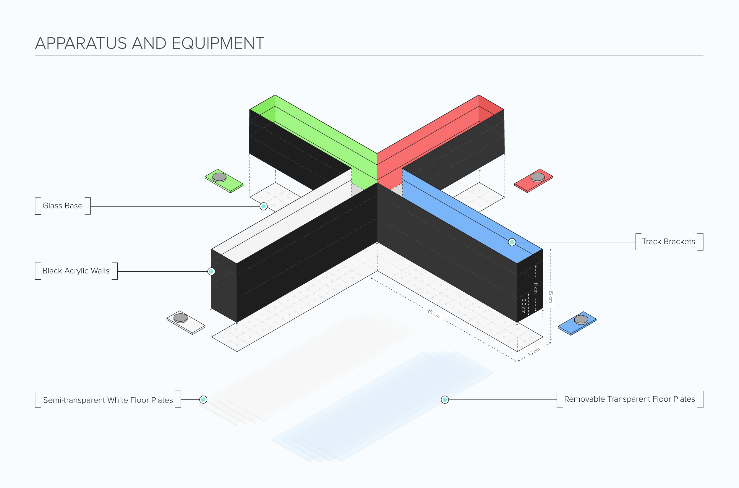
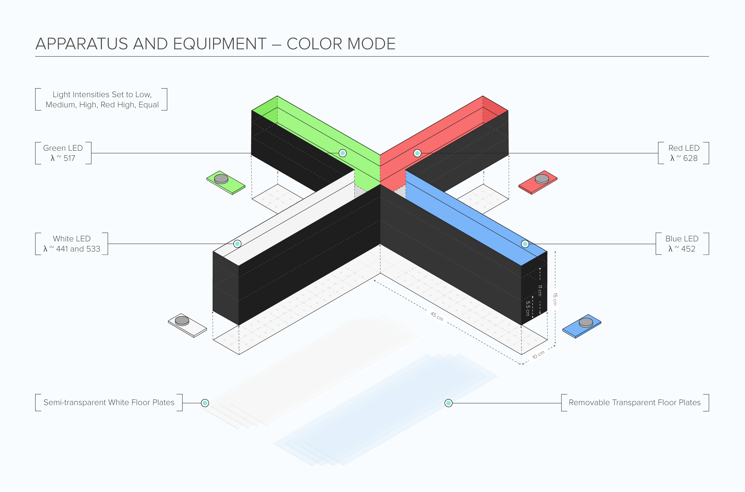
The Visual X Maze (ViS4M) apparatus is composed of black acrylic walls attached to a glass or acrylic base. Each arm of the maze measures 45 cm in length, 10 cm in width, and 15 cm in height. Transparent and semi-transparent white floor plates are placed on the bottom level of each arm of the maze using small acrylic track brackets.
Maze Engineers offers the Visual X Maze (ViS4M).
Features
Appearance
Shock Bars
Maze Mode Options
Illuminance
Price & Dimensions
Mouse Size
$ 3890
Per Month- Each arm: 45 cm total length x 10 cm total width x 15 cm total height from glass or acrylic base to top of the maze
- Floor plate insertion gaps at 6cm or 11cm above a glass base
- Central arena: 10 cm x 10 cm
- Total length from the end of the arm to the end of the arm: 100 cm
- The 90-degree angle of each maze arm to one another
Rat Size
$ 4990
Per Month- Each arm: 58 cm total length x 13 cm total width x 19 cm total height from glass or acrylic base to top of the maze
- Floor plate insertion gaps at 7.2cm or 14cm above a glass base
- Central arena: 13 cm x 13 cm
- Total length from the end of the arm to the end of the arm: 130 cm
- The 90-degree angle of each maze arm to one another
Documentation
Introduction
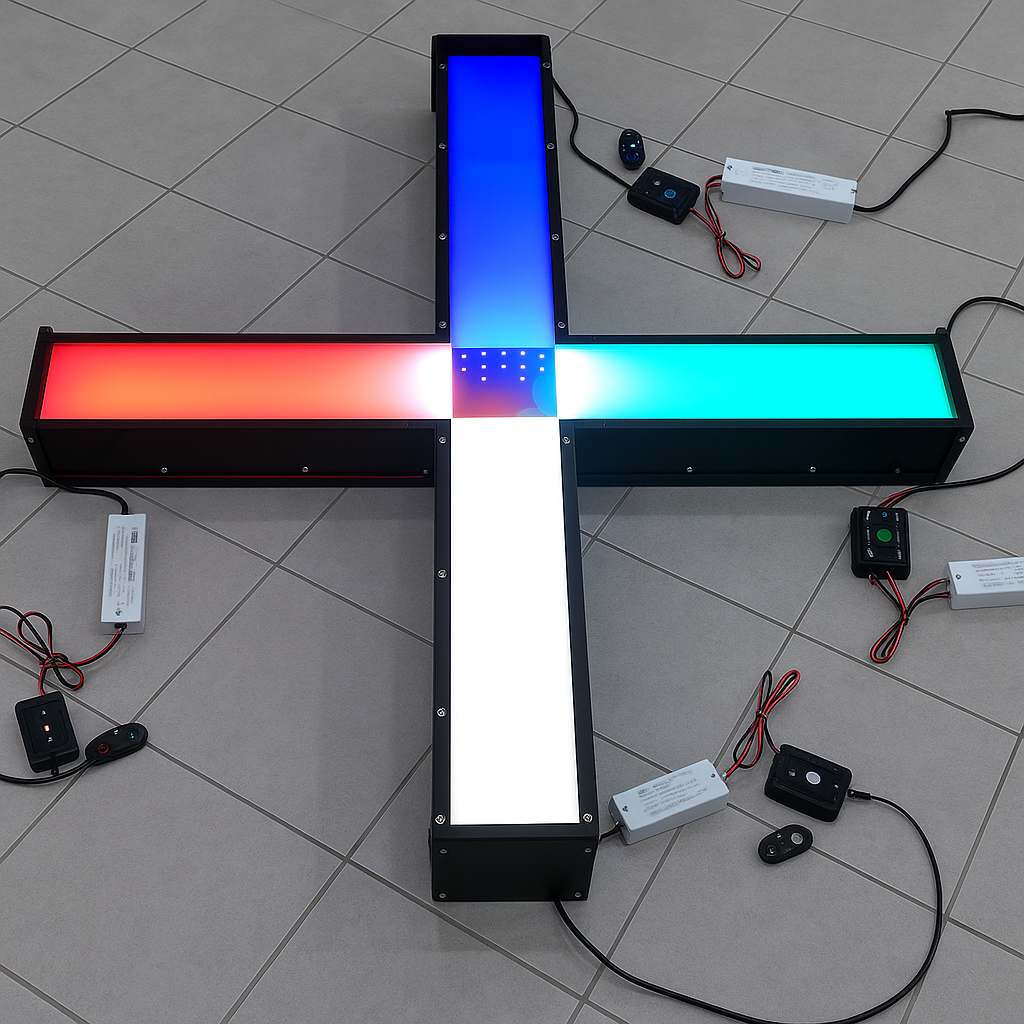
The Visual X Maze (ViS4M) is used to study visual sensitivity in rodents. It is an X-shaped apparatus that can be equipped with either light-emitting diodes (LEDs) to study color vision or different colored objects to study contrast vision in rodents (Vit et al., 2021, Vit et al., 2020).
The assessment of visual abilities is an important factor in the evaluation of visual impairments. Visual impairments, particularly those associated with amyloid plaque deposits can also indicate the development of disorders such as Alzheimer’s disease (Koronyo-Hamaoui, M., et al., 2011; Koronyo, Y. et al. 2017; Dumitrascu O., et al., 2020; Hart, N., et al., 2016; La Morgia, C., et al., 2015; Shi, H., Koronyo, Y., et al., 2020; Shi H, Koronyo Y, et al 2020a; Doustar J, et al., 2020). Deficits in vision, especially impairments in color and contrast sensitivity are common symptoms (Risacher et al., 2013; Kirby, Bandelow, & Hogervorst, 2010). The Visual X Maze allows the assessment of color vision and contrast vision in rodents, which can be used as a translational tool to study visual impairments associated with Alzheimer’s disease. During color vision tests, the maze is equipped with LEDs that emit either red, green, blue, or white light in each arm of the maze. The subject’s innate exploratory behavior is tested, and spontaneous alternations and transitions between each arm of the maze can be observed, which help indicate color preferences and color discrimination. Moreover, different light intensities can also be used to observe how they affect exploratory behaviors in the subjects.
In contrast vision testing, identical objects of different colors are utilized instead of the LEDs. The objects are placed halfway through each arm of the maze and create a contrast with the black walls and white floors of the apparatus. Apart from evaluating rodents’ visual abilities, the Visual X Maze also allows the evaluation of cognitive functions in rodents by observing revisits between the maze’s arms to assess learning and memory behaviors.
Other apparatuses used to evaluate rodents’ visual abilities include the Visual Water Box, the Visual Discrimination Chamber, the Visual Cliff Test, and the Modified Visual Cue Y Maze.
Apparatus and Equipment
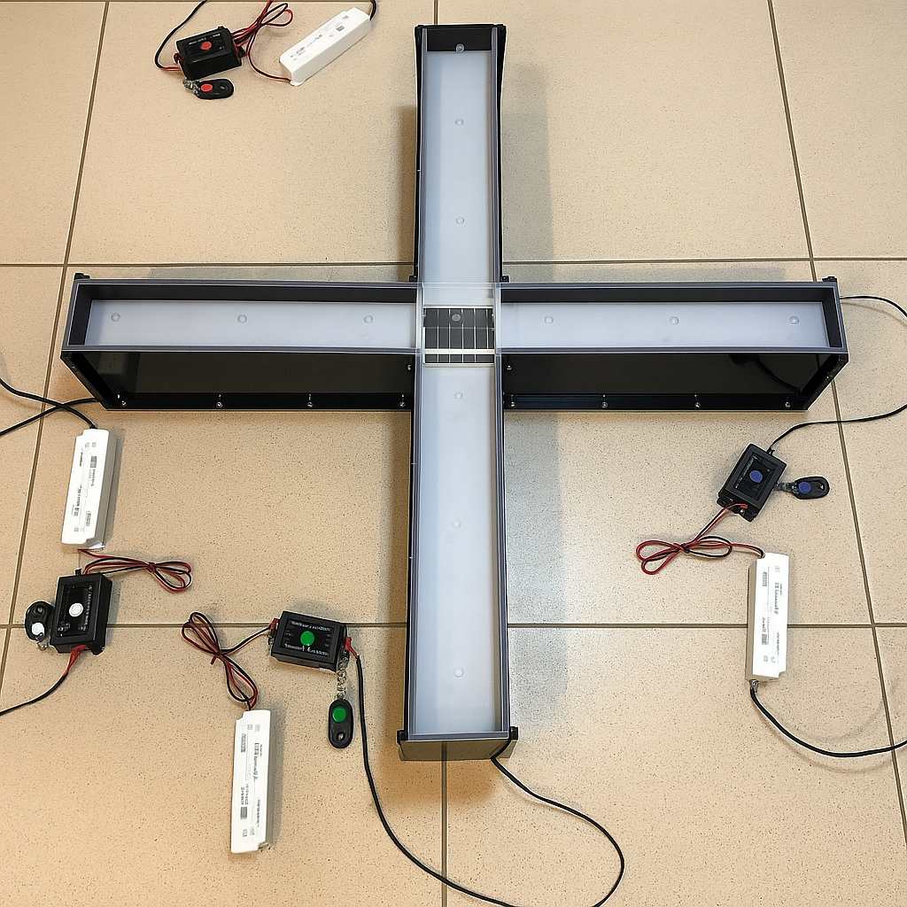
The Visual X Maze (ViS4M) is an “X” shaped apparatus that is composed of black acrylic walls attached to a glass or acrylic base. Each arm of the maze measures 45 cm in length, 10 cm in width, and 15 cm in height and is equipped with transparent ceiling covers with perforations. In each arm of the maze, removable transparent and semi-transparent white floor plates can be inserted using track brackets.
Below each floor plate, small acrylic track brackets are attached to the apparatus walls that allow floor plates to be inserted. Floor plates that come with the apparatus include four transparent floor plates and four semi-transparent white floor plates. In each arm of the maze, a light-emitting diode (LED) source can be directly inserted into the base. Each LED source consists of a strip of 40 small LEDs arranged in four lines and connected to a dimmer low-voltage transformer. The LED sources provide chromatic light at a single wavelength λ (one red source λ ~628; one green source λ ~517; one blue source λ ~452nm) or a mix of λ (~441 and 553 one white source) and can be controlled by individual remote devices.
Training Protocol
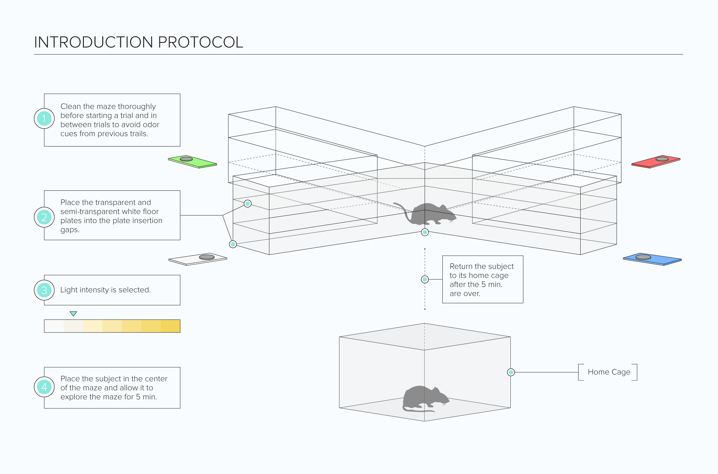
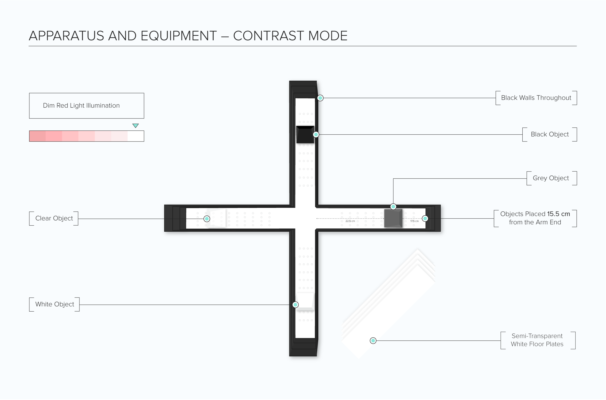
Clean the maze thoroughly before starting a trial and in between trials to avoid odor cues from previous trails. Conduct testing in a dark room with the sole light source being the maze’s illuminated arms for color mode trials. For contrast mode testing, an external dim red light illuminates the maze. A tracking and recording system such as the Noldus Ethovision XT can be used to assist with observations.
Habituation
Place the subjects in a dark testing room and allow them to habituate with the dark for 30 minutes.
Visual X Maze Color Mode Task
Place transparent and semi-transparent white floor-plates into the arm of each maze via the plate insertion gaps. Put the light intensity on low for the four arms of the maze. Place the subject in the center of the maze and allow it to explore the maze for 5 minutes. Return the subject to its home cage after the 5 minutes are over. Conduct trials over five days with the light intensity on the four following days being medium, high, red-high, and finally equal. Additionally, the subjects can be tested using a no color light condition in which the LED sources are not lit, and the assessment room dimly lit. The no color light condition can be carried out before the other light intensity conditions based on experimental needs.
Features
- 4 Light-emitting diode strips in red, blue, green, and white in color with bulbs embedded in epoxy domes
- Wavelengths λ: Red: 628, FWHM 17 nm; Green: 517, FWHM 31 nm; Blue: 452, FWHM 22 nm; White: λ1 441, FWHM 19 nm and λ2 533, FWHM 104 nm
- The white LED is made of a blue-616 emitting diode that also excites yellow-emitting phosphor – cerium doped yttrium aluminum 617 garnet (Ce: YAG-Y3Al5O12) crystals
- LED strip source consists of an array of a surface-mounted device (SMD) 3528 LED chips evenly spaced in four rows (27 LED chips per row)
- Each chip has an emission angle of 120°
- The dimmer of LEDs using pulse width modulation (PWM) technology
Visual X Maze Contrast Mode Task
Place the transparent floor plates at the bottom level with an additional semi-white transparent plate below this. Place identical objects of different shades (black, grey, clear, and white) in the middle of each arm of the apparatus. Provide the maze with red light illumination. Place the subject in the center of the maze and allow it to explore the maze for 5 minutes. Return the subject to its home cage after the 5 minutes are over. Conduct one trial before and one trial after color mode testing.
Features
- Triangular based prism objects in white, grey, black, and transparent in color
- Object dimensions (9 cm width, 5cm length, 1cm height)
- Objects placed 22.5 cm from the entrance of each arm
Literature Review
Investigation of color and contrast visual sensitivity in mice using the Visual X Maze
Vit et al investigated color and contrast visual sensitivity in aging mice and mice with Alzheimer’s disease (AD) using the Visual X Maze (ViS4M) (Vit et al., 2021, Vit et al., 2020). Cohorts of male and female wild-type (WT) mice and double-transgenic APPSWE/PS1ΔE9 mouse models of AD (AD+ mice) of 8.5, 13, and 18-months old were used in the study. The subjects were first tested for color vision changes or impairments using color mode. LEDs placed in the base of the maze provided light at the following wavelengths (λ): red (λ ~628nm), green (λ ~517nm), blue (λ ~452nm), or mixed λ for white light. The 8.5-month old mice were also tested in a no color condition in which the LED sources were not lit. Various combinations of mesopic and photopic illuminations were also tested that included low, medium, high, red-high, and equal conditions. Spontaneous alternations between the arms of the maze were observed, and results indicated that the WT mice tended to consecutively enter each arm of the maze before revisiting any arm. In no color condition, it was observed that alternations were substantially higher than chance probability. However, alterations were lower in the no color condition as compared to red-high and equal conditions. In the AD+ mice, it was observed that alternations were around the chance level in all ages and all light intensity conditions. The percentage of alternations (% alterations) was substantially reduced in AD+ mice that progressed with aging, compared to the WT mice that had a constant % alternation with aging. Further results indicated that the subjects from both experimental groups spent more time in the red arm of the maze and the least amount of time in the blue arm. At 13-months of age, the AD+ mice spent significantly less time in the white arm as compared to the WT mice. A percentage of unidirectional and bidirectional directions between the arms of the maze were also analyzed. An increase in percentage transitions was observed from the green to red arms and green to blue arms, specifically in the 8.5-month old AD+ mice. Moreover, there was a substantial decrease from white to black and black to white transitions under the red-high and equal conditions as compared to the no-color condition. The AD+ mice also had higher blue to green and green to blue transitions under red-high and equal conditions as compared to the WT mice.
The subjects were then tested for contrast sensitivity by placing identical objects of different colors (white, black, grey, and clear) in the maze’s four arms. No colored lights were used. Alike to color mode tests, the WT mice spontaneously alternated between the four arms of the maze compared to the AD+ mice. The WT mice did not exhibit a preference for any arm; however, the AD+ mice had a higher preference in the arm with a clear object and a lower preference for the black arm. Observations of arm transitions revealed impaired contrast sensitivity in the AD+ mice.
The subjects were also tested using the Barnes Maze to assess their long-term memory. The results indicated that the 13-month old AD+ mice exhibited long-term memory impairment as compared to the WT mice.
Data Analysis
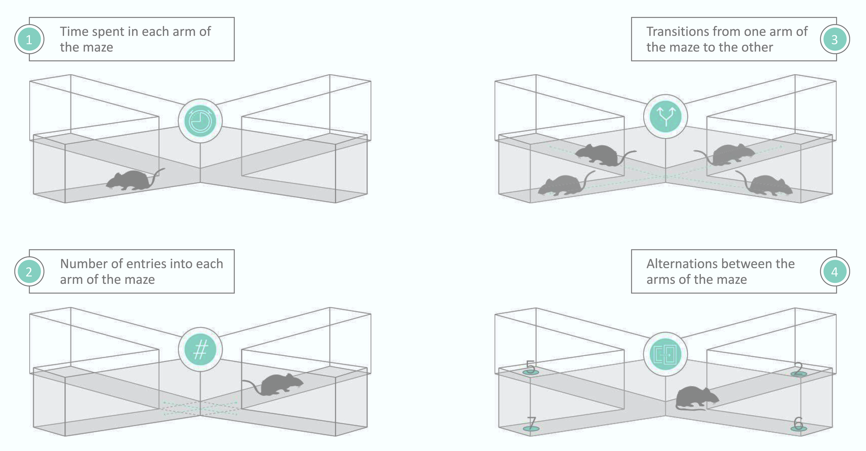
The following parameters can be observed using the Visual X Maze (ViS4M):
- Time spent in each arm of the maze
- Number of entries into each arm of the maze
- Alternations between the arms of the maze
- Transitions from one arm of the maze to the other
Strengths and Limitations
Strengths
The Visual X Maze (ViS4M) is used to study color sensitivity and contrast sensitivity in rodents. It can be equipped with different colored LEDs to study color preferences and color discrimination. Different light intensities can also be used to observe how they affect exploratory behaviors. Identical objects of different colors can also be utilized to examine contrast vision since the objects contrast with the black walls and white floors of the apparatus. Pre-training is not required using the Visual X Maze (ViS4M), so experimentation takes a short amount of time. Similar to other apparatuses such as the Y-Maze and T-maze, spontaneous alteration between the four arms of the maze can be observed. Moreover, the Visual X Maze (ViS4M), also allows the assessment of cognitive abilities in rodents by analyzing working memory through observing the number of revisits to each arm of the maze. The Visual X Maze (ViS4M) can be used as a reliable tool in exploring visual impairments in rodents, which can be used to study disorders such as Alzheimer’s disease, where visual defects are a common symptom.
- Subjects require minimal to no training to use this maze
- This novel maze specifically studies aspects of color and contrast discrimination in subjects
- Options for studying color vision or contrast vision in separate experiments
- Short experimentation time
- Minimal software requirements
- Simple experimentation protocol
Limitations
The exploratory drive of the subjects is highly important in solving the task. Factors such as age, gender, and strain of the subjects may affect task performance. The presence of odor cues from previous trials may affect task performance. Unintentional stimuli may affect the way the subject performs the task.
- It must be ensured that any unintentional stimuli are kept to a minimum to ensure accurate readings.
- Equipment must be cleaned between each subject and trial
- External stimuli such as noise must be kept to a minimum
- Factors such as age, gender, the subject strain could affect performance
Summary
- The Visual X Maze (ViS4M) is used to study visual sensitivity in rodents based on their color vision and contrast vision.
- It is an X-shaped apparatus with black walls and semi-transparent white floors. Removable floor plates can be inserted at different levels of the apparatus. LED emitters can be inserted directly into the maze base to provide different color illumination to each arm of the maze.
- Different colored objects can also be placed in the apparatus to serve as visual cues.
- The Visual X Maze (ViS4M) allows the evaluation of color preferences in rodents as well as color discrimination. Moreover, the working memory of the subjects can also be analyzed.
References
- Vit, J.P., Fuchs, D.T., Angel, A., Levy, A., Lamensdorf, I., Black, K., Koronyo, Y., Koronyo-Hamaoui. M. (2020). Color and contrast vision in mouse models of aging and Alzheimer’s disease using a novel visual-stimuli four-arm maze. Sci Rep 11,1255 (2021). https://doi.org/10.1038/s41598-021-80988-0
- Vit, J., Fuchs, D., Angel, A., Levy, A., Lamensdorf, I., Black, K. L., Koronyo, Y. and Koronyo-Hamaoui, M. (2021). Visual-stimuli Four-arm Maze test to Assess Cognition and Vision in Mice. Bio-protocol 11(22): e4234. DOI: 10.21769/BioProtoc.4234.
- Kirby, E., Bandelow, S., & Hogervorst, E. (2010). Visual impairment in Alzheimer’s disease: a critical review. Journal of Alzheimer’s disease: JAD, 21(1), 15–34. https://doi.org/10.3233/JAD-2010-080785.
- Risacher, S. L., Wudunn, D., Pepin, S. M., MaGee, T. R., McDonald, B. C., Flashman, L. A., Wishart, H. A., Pixley, H. S., Rabin, L. A., Paré, N., Englert, J. J., Schwartz, E., Curtain, J. R., West, J. D., O’Neill, D. P., Santulli, R. B., Newman, R. W., & Saykin, A. J. (2013). Visual contrast sensitivity in Alzheimer’s disease, mild cognitive impairment, and older adults with cognitive complaints. Neurobiology of Aging, 34(4), 1133–1144. https://doi.org/10.1016/j.neurobiolaging.2012.08.007.
- Koronyo-Hamaoui, M., et al. (2011). Identification of amyloid plaques in retinas from Alzheimer’s patients and noninvasive in vivo optical imaging of retinal plaques in a mouse model. NeuroImage, 54(1),
204-217. https://doi.org/10.1016/j.neuroimage.2010.06.020 - Koronyo, Y. et al. (2017). Retinal amyloid pathology and proof-of-concept imaging trial in Alzheimer’s disease. JCI Insight. 2017 Aug 17;2(16):e93621. https://doi.org/10.1172/jci.insight.93621.
- La Morgia, C., et al. (2016). Melanopsin retinal ganglion cell loss in Alzheimer’s disease. Ann Neurol. 2016 Jan;79(1):90-109. https://doi.org/10.1002/ana.24548
- Hart, N., et al. (2016). Ocular indicators of Alzheimer’s: exploring disease in the retina. Acta Neuropathol 132,767–787. https://doi.org/10.1007/s00401-016-1613-6
- Dumitrascu O., et al. (2020). Sectoral segmentation of retinal amyloid imaging in subjects with cognitive decline. Alzheimer’s Dement (Amst), 12(1): e12109. PMID: 33015311. https://doi.org/10.1002/dad2.12109
- Shi, H., Koronyo, Y., et al. (2020). Identification of early pericyte loss and vascular amyloidosis in Alzheimer’s disease retina. Acta Neuropathol 139, 813–836. https://doi.org/10.1007/s00401-020-02134-w
- Doustar J, et al. (2020). Parallels between retinal and brain pathology and response to immunotherapy in old, late-stage Alzheimer’s disease mouse models. Aging Cell. 19(11):e13246. https://doi.org/10.1111/acel.13246
- Shi H, Koronyo Y, et al (2020a). Retinal capillary degeneration and blood-retinal barrier disruption in murine models of Alzheimer’s disease. Acta neuropathol commun 8, 202. https://doi.org/10.1186/s40478-020-01076-4
- Gaire, B. P., Koronyo, Y., Vit, J. P., Hutton, A., Subedi, L., Fuchs, D. T., Swerdlow, N., Rentsendorj, A., Shahin, S., Martinon, D., Robinson, E., Ljubimov, A. V., Schneider, J. A., Schneider, L. S., Hawes, D., Graham, S. L., Gupta, V. K., Mirzaei, M., Black, K. L., . Koronyo-Hamaoui, M. (2026). Identification of Chlamydia pneumoniae and NLRP3 inflammasome activation in Alzheimer’s disease retina. Nature Communications, 17(1), 771. https://doi.org/10.1038/s41467-026-68580-4
- Weekman, E.M., Rogers, C.B., Sudduth, T.L. et al. Hyperhomocysteinemia-induced VCID results in visual deficits, reduced neuroinflammation and vascular alterations in the retina. J Neuroinflammation 22, 23 (2025). https://doi.org/10.1186/s12974-025-03332-7
Are you an academic scientist and creator?
Learn how your lab can tech transfer this and similar devices from your lab to industry.