
- Arena: Acrylic open field without walls, 140 × 110 cm; clear
- Transparent acrylic floor (0.6 cm)
- Underside of floor: Unity-gain Dual Vision Fabric screen stretched over a second acrylic sheet (150 × 110 × 1.25 cm) for rear projection with a short-throw projector
- Project available on request
Investigating visual cognition through spatially projected environments
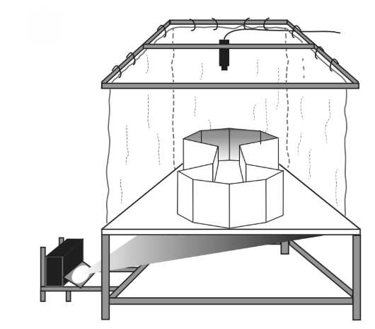
The Floor Projection Maze is a modular apparatus developed to study visual information processing, learning, memory, and attention in rodents. Originally described by Burwell et al. (2014), it provides a controlled environment for investigating visual cognition through automated tasks.
Visual cues are projected onto a semi-transparent floor illuminated from below by a digital projector. This configuration aligns with rodents’ natural downward gaze, producing a biologically relevant and immersive testing environment.
The maze’s adaptable design supports rapid modification of experimental conditions, allowing researchers to test a wide range of behavioral paradigms.
Price & Dimensions
Rat Floor Maze
$ 4990
Per MonthApparatus & Equipment
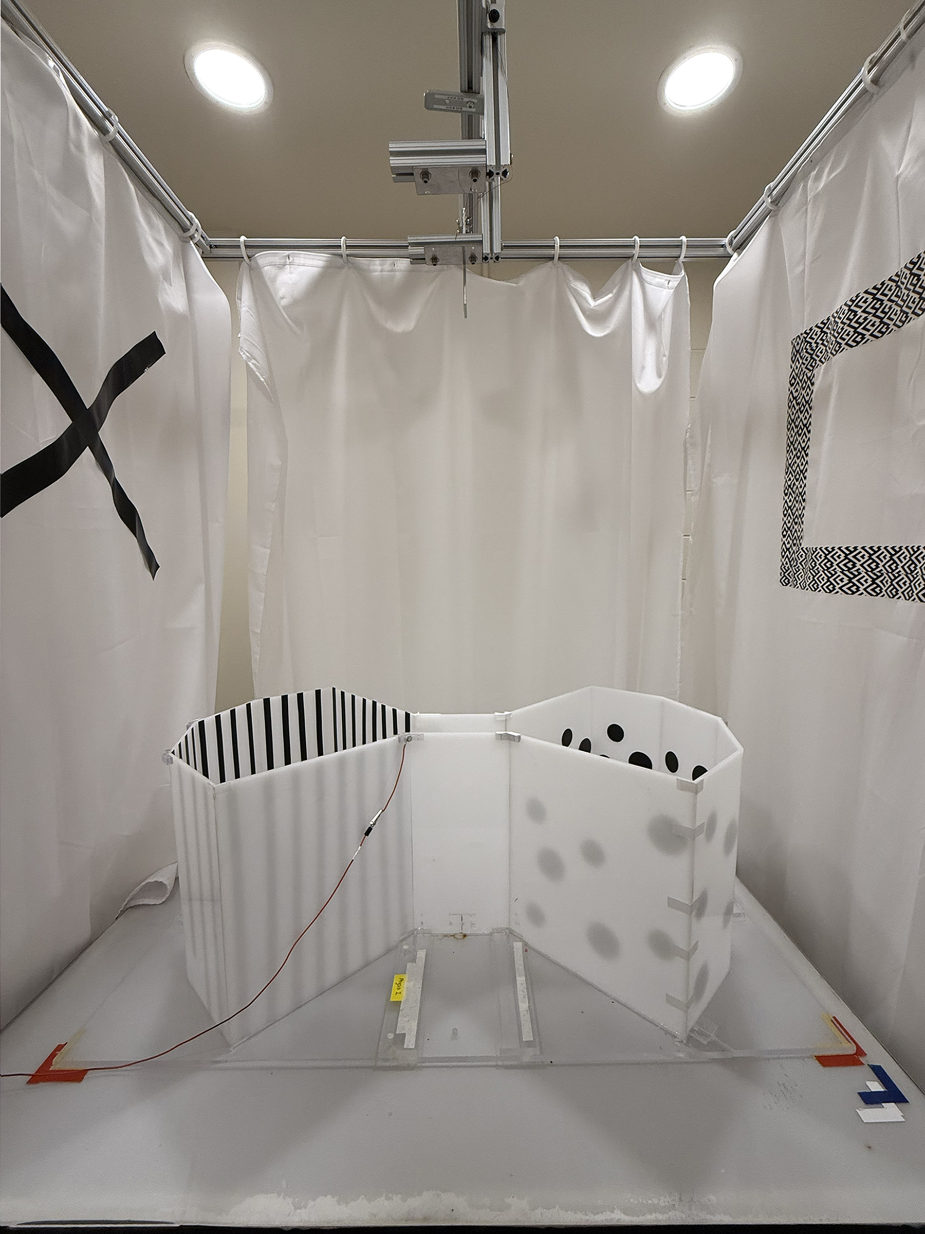
Overhead Camera & Tracking
Mounted above the arena, the camera provides continuous video for position and trajectory reconstruction. When paired with ConductVision tracking, coordinates stream to the control software in real time.
Arena Surface (Clear Acrylic Floor)
Subjects move on a clear acrylic plate that serves as the behavioral arena while allowing visual stimuli to be projected from underneath.
Digital Projector + Mirror
A calibrated projector directs images onto a first‑surface mirror set at a fixed angle. The mirror reflects the stimuli to the underside of the floor, producing sharp, high‑contrast patterns with minimal parallax.
Reward Delivery (ICS & Alternatives)
For operant tasks, intracranial stimulation to the medial forebrain bundle can be delivered as an immediate reinforcer. The system also supports lickometers and pellet dispensers when consumable rewards are preferred.
Control Software (Automation & Integration)
A single computer coordinates stimulus presentation, tracking input, and reward timing, enabling fully automated sessions and reducing experimenter bias while increasing trial throughput.
Data Analysis
Behavioral Performance Analysis
Behavioral outcomes were assessed through accuracy, reaction times, and error rates across visual biconditional discrimination (vBCD) and visuospatial attention (VSA) tasks. Learning curves were generated to measure performance improvements across sessions, quantifying accuracy and response efficiency over time.
Reaction latency was recorded from stimulus onset to target entry, while omission and perseverative errors provided indicators of attention and behavioral control.

Spatial & Trajectory Analysis
Path tracking and movement metrics were derived from overhead video recordings. Trajectories were reconstructed to visualize exploration strategies and navigation efficiency. Dwell time within predefined areas (e.g., Ready or Image Zones) quantified spatial preferences.
Exploration was further characterized by path length, speed, and thigmotaxis (tendency to remain near walls), providing insight into anxiety and motivation levels. Approach bias (left vs. right) was analyzed across trials to detect spatial decision tendencies.

Stimulus-Dependent Behavior
Context-dependent decision-making was analyzed to determine how floor pattern context altered image selection in vBCD paradigms. In visuospatial attention tasks, accuracy and speed were examined as a function of stimulus distance and target position.
Cue salience tests evaluated how variations in brightness, size, or contrast influenced response probability, reflecting perceptual sensitivity and adaptive learning.
Neural & Electrophysiological Analysis
(applied when simultaneous neural recording is used)
Neural activity was recorded using spike sorting to identify single units and analyze firing rate changes during stimulus presentation, decision-making, or reward delivery. Event-related firing patterns were aligned to trial events such as stimulus onset, choice, or reward to map temporal correlations.
Local field potentials (LFPs) were analyzed to study oscillatory dynamics related to attention, memory, and learning. When multi-site recordings were available, cross-region coherence and correlation analyses were used to characterize network-level interactions.
Training Protocol
Early Shaping
The early shaping phase focuses on exploration and familiarization with key zones of the maze. Over the course of five days, subjects learn to associate specific areas with rewards through incremental exposure.
- Day 1: Habituate to the testing room for 10 minutes (equipment on).
- Day 2: Habituate to the arena for 10 minutes.
- Day 3: Connect ICS and headstage tethers; 10-minute arena session.
- Day 4: Adjust ICS amplitude (20–80 µA) via informal place-preference testing.
- Day 5: Deliver ICS to associate the Ready Area and Image Areas with reward until alternating behavior develops.

Intermediate Shaping
During intermediate shaping, animals learn to maintain a “ready position.” Trials begin with a 50 dB white noise cue, which turns off once the subject enters the Ready Area. Automated ICS rewards reinforce successful entries and holds.
Reward probability is progressively reduced to 5–10% for stable responses. Hold durations start around 200 ms and increase in 100 ms increments, reaching up to 1,200 ms as the animal’s performance stabilizes.
Late Shaping (Task-Specific Automation)
The late phase focuses on full automation of task-specific paradigms, including visual biconditional discrimination (vBCD) and visuospatial attention (VSA) tasks.
vBCD: Trials begin with a white-noise cue and randomized ready latency (700–1,200 ms). Subjects view paired images and receive rewards for correct zone entries. Incorrect responses trigger correction trials. Once simple discrimination is learned, distinct floor patterns are added to create context-dependent rules.
VSA: Random ready-latency (1,000–1,600 ms). Following a successful hold, one circle location is briefly illuminated; entry into the target zone is rewarded. Illumination duration decreases as accuracy improves, transitioning from 5 s to 500 ms. Incorrect or omitted trials receive no reward, and the next trial begins from the opposite side.

Apparatus
The arena consists of an open field with a transparent acrylic floor (0.6 cm thick), layered with a unity-gain Dual Vision Fabric screen for rear projection via a short-throw projector. The setup includes an overhead camera for real-time tracking and behavioral recording.
A matte-white acrylic wall structure (45–50 cm height) defines the arena boundaries. The example configuration uses a double-sided bowtie arena with four software-defined areas: East Image, West Image, East Trial-Ready, and West Trial-Ready zones.
Visual cues are projected using custom software paired with the Dual Vision screen. ICS rewards are delivered via bipolar square-wave stimulation (pulse width 500 µs, delay 500 µs, 100 Hz) controlled by PC software or a manual button box when needed.
Animals & Handling
Experiments use naïve male Long-Evans rats (~P22). Animals are pair-housed for one week, then handled for 5 minutes daily. Food scheduling begins at 250–275 g to maintain 85–90% free-feeding weight, increasing target weight by 10 g/month until reaching 350 g. Single housing begins at least one week before surgery.
Electrode implantation is performed under isoflurane anesthesia. Craniotomies are made after locating bregma and lambda. The ICS electrode is implanted into the medial forebrain bundle (MFB) at AP −2.7 mm, ML ±1.8 mm, DV −8.5 mm. A backup ICS electrode may be implanted contralaterally.
Electrodes are secured using bone cement, ensuring the ICS pedestal remains uncapped. Recording electrodes are implanted at the target site and stabilized with cement. Animals recover for at least seven days before beginning experiments.
Literature Review
Functional Differentiation of Dorsal and Ventral Posterior Parietal Cortex of the Rat: Implications for Controlled and Stimulus-Driven Attention
Cerebral Cortex, 2022;32: 1787–1803
Yang, Dokovna, and Burwell (2022) used the Floor Projection Maze to investigate functional differentiation in the rat posterior parietal cortex (PPC). By combining anatomical tract-tracing with simultaneous electrophysiological recordings during a visuospatial attention (VSA) task, the study found clear evidence for division of labor within PPC. Anatomical data showed that the ventral PPC (VPPC) had stronger reciprocal connections with the postrhinal cortex (POR) and received preferential input from specific thalamic subdivisions, suggesting a role in bottom–up processing. Electrophysiological analyses revealed that VPPC neurons responded more rapidly and in greater numbers to stimulus onset compared to dorsal PPC (DPPC), indicating a specialization for stimulus-driven (bottom–up) attention. In contrast, DPPC neurons were more engaged in task-related signals and top–down control, supporting perception-to-action processes. Together, these results provide the first evidence that dorsal and ventral PPC in rats are functionally distinct, with DPPC mediating controlled, top–down attention and VPPC mediating fast, stimulus-driven attention.
Neuronal Activity in the Rat Pulvinar Correlates with Multiple Higher-Order Cognitive Functions
Yang & Burwell (2020) used the Floor Projection Maze to record neuronal activity in the rat pulvinar (lateral posterior thalamus) during a visuospatial attention (VSA) task. Rats monitored three possible stimulus locations, and their neural responses were analyzed across key behavioral epochs (stimulus onset, target selection, reward approach).
The findings showed that over three-quarters of pulvinar neurons (74–79%) exhibited task-related activity, demonstrating that this thalamic nucleus participates in multiple higher-order cognitive processes. Specifically:
-
Stimulus-driven and controlled attention: Some neurons increased firing immediately after stimulus onset (bottom–up attention), while others showed activity during pre-stimulus monitoring (top–down attention).
-
Decision-making: A significant subset of cells differentiated correct vs. incorrect target selections, indicating involvement in guiding choice behavior.
-
Reward processing: Many neurons signaled trial outcomes during the reward-approach phase, encoding success or failure.
-
Spatial reference frames: About 37% of cells encoded allocentric (east vs. west side) location and 30% encoded egocentric (left, center, right) position; importantly, some cells showed mixed selectivity for both frames, suggesting pulvinar’s role in translating spatial information across reference systems.
References
- Jacobson TK, Ho JW, Kent BW, Yang FC, Burwell RD. Automated visual cognitive tasks for recording neural activity using a floor projection maze. J Vis Exp. 2014 Feb 20;(84):e51316. doi: 10.3791/51316. PMID: 24638057; PMCID: PMC4130232.
-
Yang FC, Dokovna LB, Burwell RD. Functional Differentiation of Dorsal and Ventral Posterior Parietal Cortex of the Rat: Implications for Controlled and Stimulus-Driven Attention. Cereb Cortex. 2022 Apr 20;32(9):1787-1803. doi: 10.1093/cercor/bhab308. PMID: 34546356; PMCID: PMC9070340.
-
Yang FC, Burwell RD. Neuronal Activity in the Rat Pulvinar Correlates with Multiple Higher-Order Cognitive Functions. Vision (Basel). 2020 Mar 1;4(1):15. doi: 10.3390/vision4010015. PMID: 32121530; PMCID: PMC7157601.